DICOM Annotation Tool for Medical Imaging
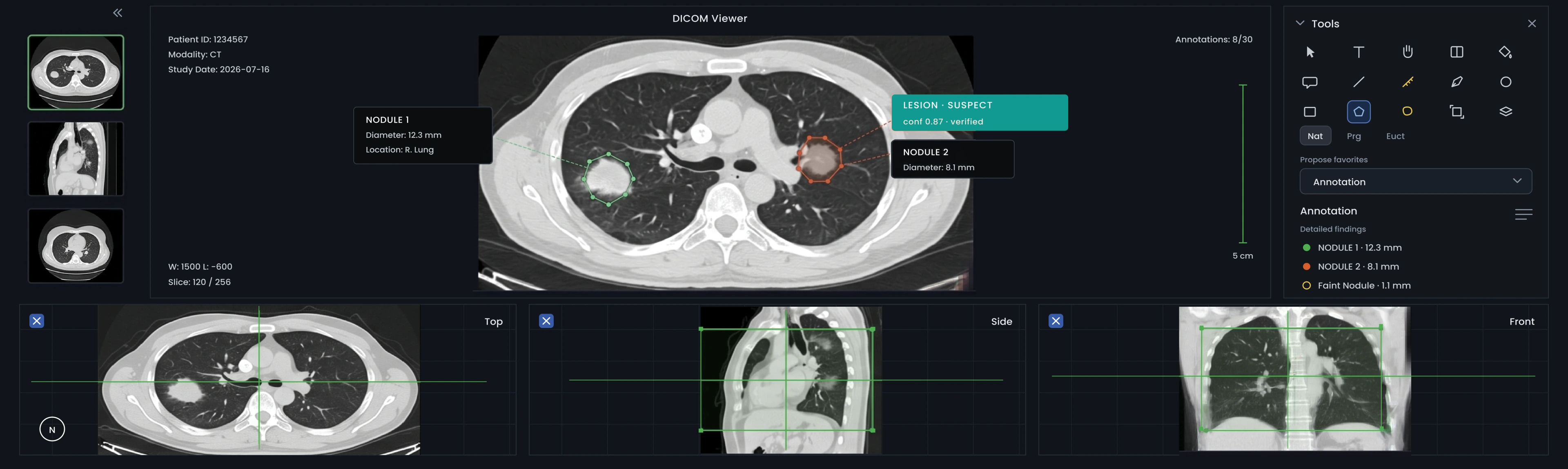
LabelFort natively annotates DICOM series, including CT, MRI, X-ray, and PET, using an Orthanc-backed medical viewer. Features include structured finding forms, measurements (length, rectangle, and ellipse ROI), and landmark annotations, with window and level control and multi-slice navigation, all under HIPAA-aligned access controls. As both a DICOM annotation tool and managed data annotation service, LabelFort offers clinical review by trained medical teams and exports every study with its audit evidence.
Training data that stands up to clinical validation & regulatory review
Clinical validation requires more than accuracy. Reviewers, such as radiologists or Notified Bodies, assess not only correct labeling but also who performed the annotation, their qualifications, the version of the finding criteria used, and whether records can withstand independent scrutiny. A DICOM annotation tool that lacks reviewer identity, versioned criteria, or per-study audit trails may produce accurate data but still fail validation. LabelFort’s clinical validation support ensures that QMS documentation, reviewer credentials, criteria versions, and per-study sign-off are generated as part of the annotation process, eliminating the need for separate, retrospective documentation.
Medical imaging annotation across modalities
Medical imaging annotation requires strict evidence standards, including PHI handling, clinical reviewer qualifications, and per-study traceability. LabelFort natively supports DICOM, series-based batches, orientation markers, and image measurement readouts, all under HIPAA-aligned access controls with de-identification. The same governed annotation pipeline applies to CT, MRI, X-ray, and PET series, so multi-modality programs do not need separate vendors or evidence standards.
How LabelFort implements clinical governance
Annotation teams are trained in clinical taxonomies and operate under radiologist or specialist supervision as part of the managed service, rather than relying on general annotators. IAA is reported per cohort, and platform audit logs record every access and action, providing the documentation required for clinical validation and QMS. Reviewer qualifications, board certification, subspecialty, and years of relevant practice are documented and attached to each record, ensuring QMS auditors receive verified credentials rather than a post hoc roster.
De-identification is performed before annotation reaches a reviewer, when permitted by the use case. The same immutable logging system tracks both labeling actions and access to the underlying study, so questions about PHI access and annotation quality are answered from a single audit trail, eliminating the need to reconcile separate systems.
Medical imaging capabilities
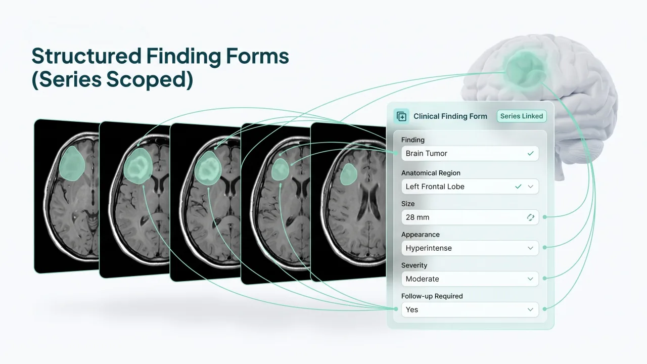
Structured Finding Forms (Series Scoped)
Structured finding forms in LabelFort are series scoped, not image scoped. This approach preserves clinical continuity, allowing findings to reference their position and appearance across multiple slices within a series. Measurements or landmarks taken on one slice remain linked to the same finding as it is tracked throughout the series.
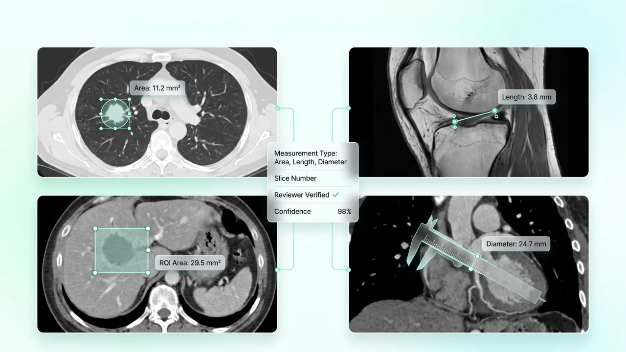
Measurements: Length, Rectangle ROI, Ellipse ROI
Length measurements record a distance directly on the image, a lesion's longest axis, a vessel diameter, the kind of number a radiology report actually cites. Rectangle and ellipse ROI capture an area rather than a single line, used when a region's extent matters more than one dimension, a mass's boundary, a zone of reduced density. All three carry the same reviewer identity and version history as every other geometry in LabelFort's DICOM annotation tool.
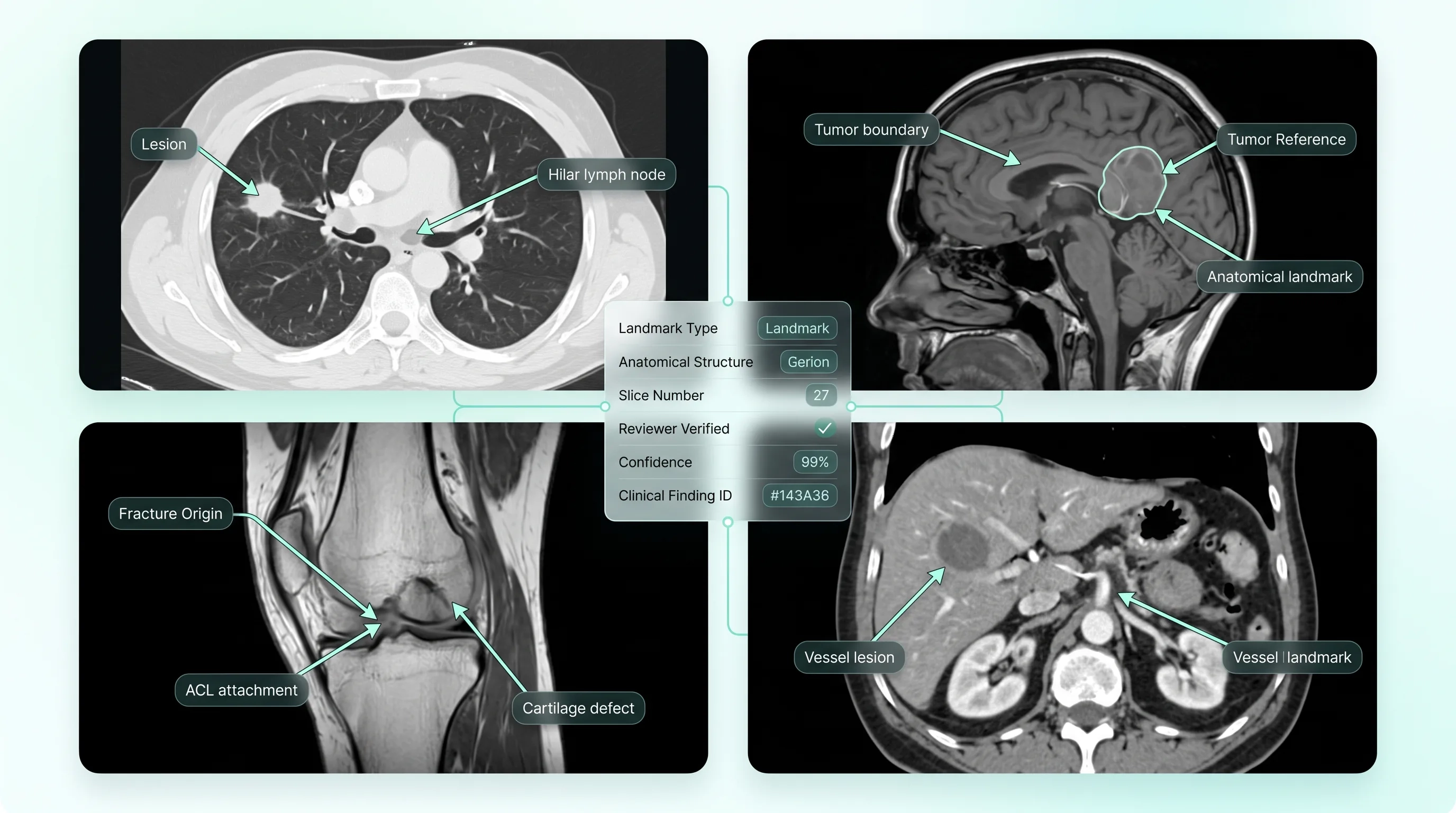
Landmark (Arrow) Annotation
An arrow landmark points to a specific finding without outlining it, the right choice when a finding's location matters more than its precise boundary, a fracture line's origin, a fixed anatomical reference point. It is the fastest annotation type available, and using it instead of a full measurement or region keeps the record honest about how precisely a finding was actually characterized.
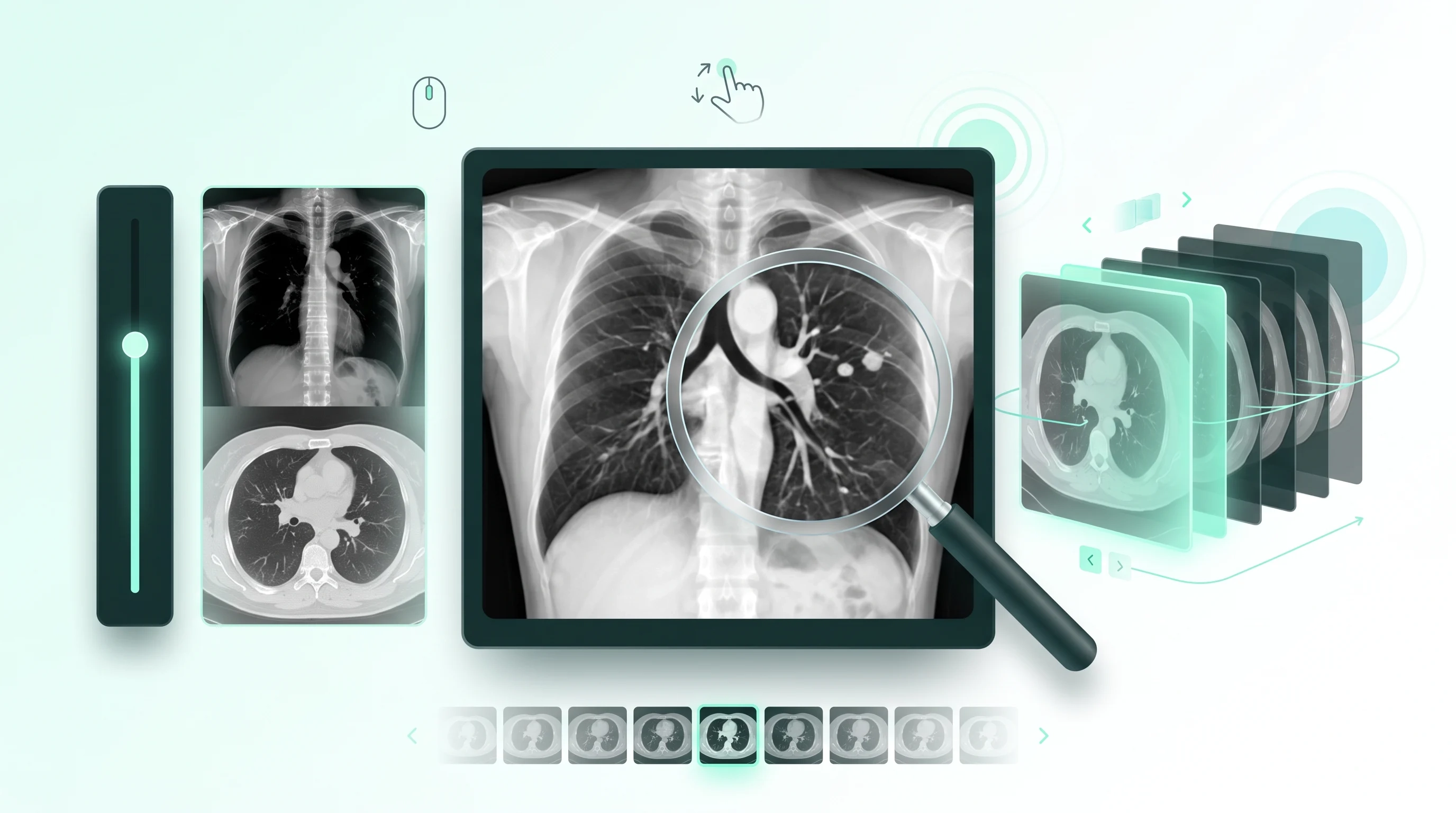
Window & Level, Zoom, & Slice Navigation
A DICOM image carries more dynamic range than a screen can show at once, window and level control lets a reviewer adjust which part of that range is visible, bone detail versus soft tissue, without altering the underlying pixel data. Zoom and multi slice navigation let a reviewer move through a series the way it's actually read clinically, not as a single flat image, which matters for medical imaging annotation work that depends on continuity across slices.
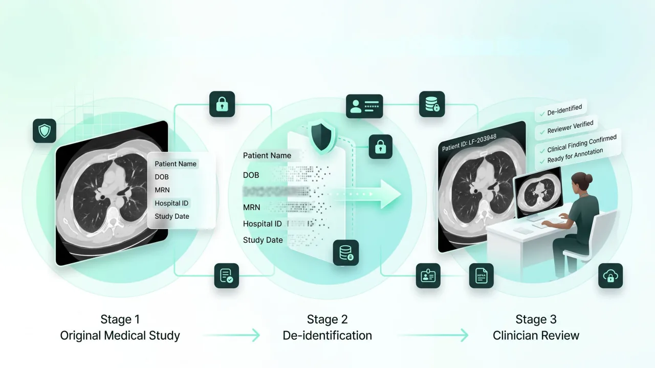
De-identification Workflows & Clinician Review in the Managed Service
De-identification runs before a study reaches a reviewer wherever the use case permits, so PHI exposure is minimized before annotation begins rather than addressed afterward. Clinician review inside the managed service means the person confirming or correcting a finding has the clinical background to recognize what the guideline is actually asking them to distinguish, not a general annotator working from a glossary.
FAQs
Is LabelFort suitable for radiology AI training data?
Yes. Native DICOM tooling (findings, measurements, landmarks), clinician review within the managed service, HIPAA-aligned controls, and per-study audit evidence make LabelFort well-suited to radiology and broader medical imaging programs.
How is PHI protected during annotation?
De-identification workflows, role-based access with purpose limitation, hardened access controls, and an immutable log of every access and action, under an ISO 27001:2022 certified ISMS.
Can this support a QMS submission or clinical validation study directly?
Yes, that's the specific gap this page is built to close. Reviewer credentials, finding criteria versions, and per-study sign-off export in the same evidence bundle as the labels, so the documentation a QMS file or validation study needs doesn't have to be reconstructed separately from what the annotation team can actually attest to.
Does this work across imaging modalities, or is it CT and MRI-specific?
The same governed pipeline covers CT, MRI, X-ray, and PET series natively, with the same de-identification, review, and audit standard applied regardless of modality.
Related services
Image Annotation
Enhanced precision and reliability for vision based models, supported by comprehensive evidence documentation.
Read More
Video Annotation
Better recall for dynamic events, with continuity that holds across every frame.
Read More
Text Annotation
Robust datasets for stable and accurate language models.
Read More
Make it provable.
Start with the free one-hour Compliance Review, your risk surface mapped to controls, a documented gap analysis, & a scoped evidence-grade PoC. Before any data moves.
Certifications & readiness